We are interested in :-
- Fluids of the Inner Ear, perilymph and endolymph.How they are maintained and regulated.
- Barriers of the Inner Ear, controlling how substances enter, leave and distribute in the ear
- Local Drug Delivery to the Inner Ear, optimizing methods to get drugs into the ear
- Computer Simulations of Drug Distribution in the Ear, using the basic physics of how drugs spread
Illustrations on the top banner are:- (Left) Our first ever 3D view of the inner ear's fluid spaces from an experiment we did back in the 1980's. The perilymphatic spaces of a guinea pig inner ear were filled with orange latex and the endolymph spaces with dark blue latex. The bone surrounding the cochlea was then dissected away. (Middle) Digital 3D reconstruction of the fluid spaces of the guinea pig ear generated from virtual slices obtained with an optical sectioning technique. Yellow:scala tympani; Green: scala vestibuli; Blue:endolymph; Cyan: round window; Brown: stapes. These data allow us to accurately quantify the dimensions of each of the fluid and tissue spaces of the ear for use in computer models. (Right) A conventional mid-modiolar histologic section through the guinea pig ear, showing the membranous and cellular structures inside. The nerve is maroon and the bone is purple.
Changes from NIH and the FDA Regarding Developing Drug Therapies
New guidelines have been introduced by NIH and the FDA to expand innovative, human-based science while reducing animal use in research. This involves using cutting-edge alternative non-animal research models, including computational models which simulate complex biological human systems, disease pathways and drug interactions.
Read how Turner Scientific is in a unique position to help you navigate these changes, through the use of FluidSim, the only sophisticated simulation of drug dispersal in the inner ear, and only available through Turner Scientific.
Analysis of Cerebrospinal Fluid (CSF) and brain tissues
Turner Scientific now offers Cerebrospinal Fluid and brain tissue testing of pharmaceuticals in a variety of species. Sampling uses state-of-the-art "clean" sampling techniques to collect high-purity samples. Click for more details.
Recent Publications
 Click for PubMed access
 Click for free access from the Frontiers website
The Turner Library
A Collection of Educational Articles Relevant to Inner Ear Drug Delivery
1) Evaluation of Candidate Drugs for Local Delivery to the Ear
2) Direct Drug Injections into Perilymph *
3) Perilymph Sampling *
5) Dilution of Perilymph Samples for Storage and Analysis
6) Background Solutions for Drugs Injected into Perilymph *
7) How can the FluidSim Cochlear Fluids Simulator help me?
8) Toxicology Assessments for Hearing and Balance *
9) "Pipeline" Services Available from Turner
* indicates downloadable/printable/sharable pdf available
Click to access available pdf files
The FluidSim Page
Inner Ear Fluids Simulation Program - Version 5.02 (August 2025)

The FluidSim program saves costs and animal usage through the three R's, which stand for Replacement, Reduction and Refinement.
Replacement: In some cases simulation of a pharmacokinetic study is sufficient to show that use of a specific drug in a specific application will either be successful or unsuccessful, making the use of animal experiments unnecessary.
Reduction: Simulations of the planned pharmacokinetic study allow the experiment to be better designed, allowing data collection time points and drug dosing amounts to be optimized, avoiding unnecessary experiments.
Refinement: Analysis of measured data with FluidSim allows the maximum value to be extracted from the collected data and the experimental design to be refined over time.
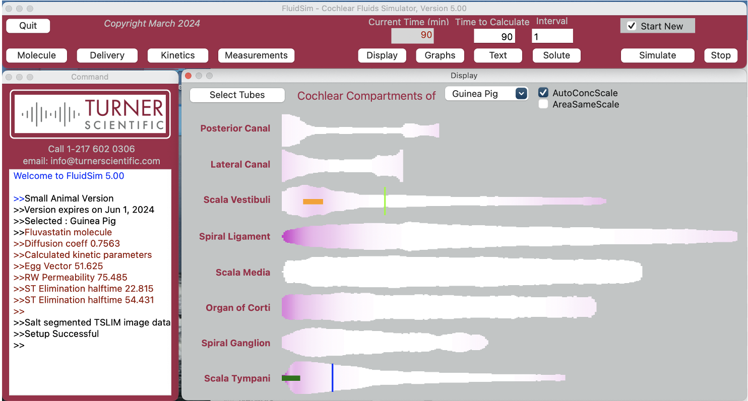
FluidSim software simulates all common drug delivery and perilymph sampling protocols used in the ear. The predictions are used to help design pharmacokinetic studies and to interpret drug measurements by accurately replicating how drugs distribute along the ear with time. Simulations can be performed for the ears of 7 animal species, including guinea pigs, mice, rats, cats, sheep, domestic pigs, cynomolgus monkeys, and the human ear.
You either download the program for yourself or we can provide assistance and do the calculations for you. If you are interested in contracting with Turner to provide computer predictions or analysis of data you have already collected, contact
