The Function of the Inner Ear
In 1861 Prosper Meniere fundamentally changed our understanding of the ear and was ostracized for it.
This summary is based on the content of rare otolaryngology textbooks available in Dr. Richard Chole’s collection at Washington University and of other vintage texts available through Google Books Advanced Search. During the 19th century there were major advances in understanding of how the ear worked. Meniere's true contribution is often overlooked.
A modern understanding of the inner ear was summarized (Cummings, Otolaryngology Head and Neck Surgery, 5th Edition, 2010) as: “The two important functions of the ear are hearing and balance. The portion of the inner ear that deals with hearing is the cochlea and the portion of the inner ear that deals with balance is collectively known as the vestibular organs (semicircular canals, utricle and saccule).”
But it wasn’t always this way.
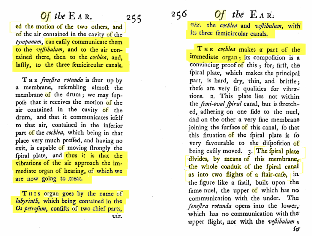
In 1734, the consensus view was that the ear was entirely the “Hearing Organ”. Under this view, different parts of the ear performed different analyses of the sound.
The Semi-circular canals coded the directionality of sound and provided enhancement or attenuation of auditory stimuli
The Saccule and Utricle coded the hearing of noises
The Cochlea coded the hearing of tones
From Thomson 1734: (images of the original texts)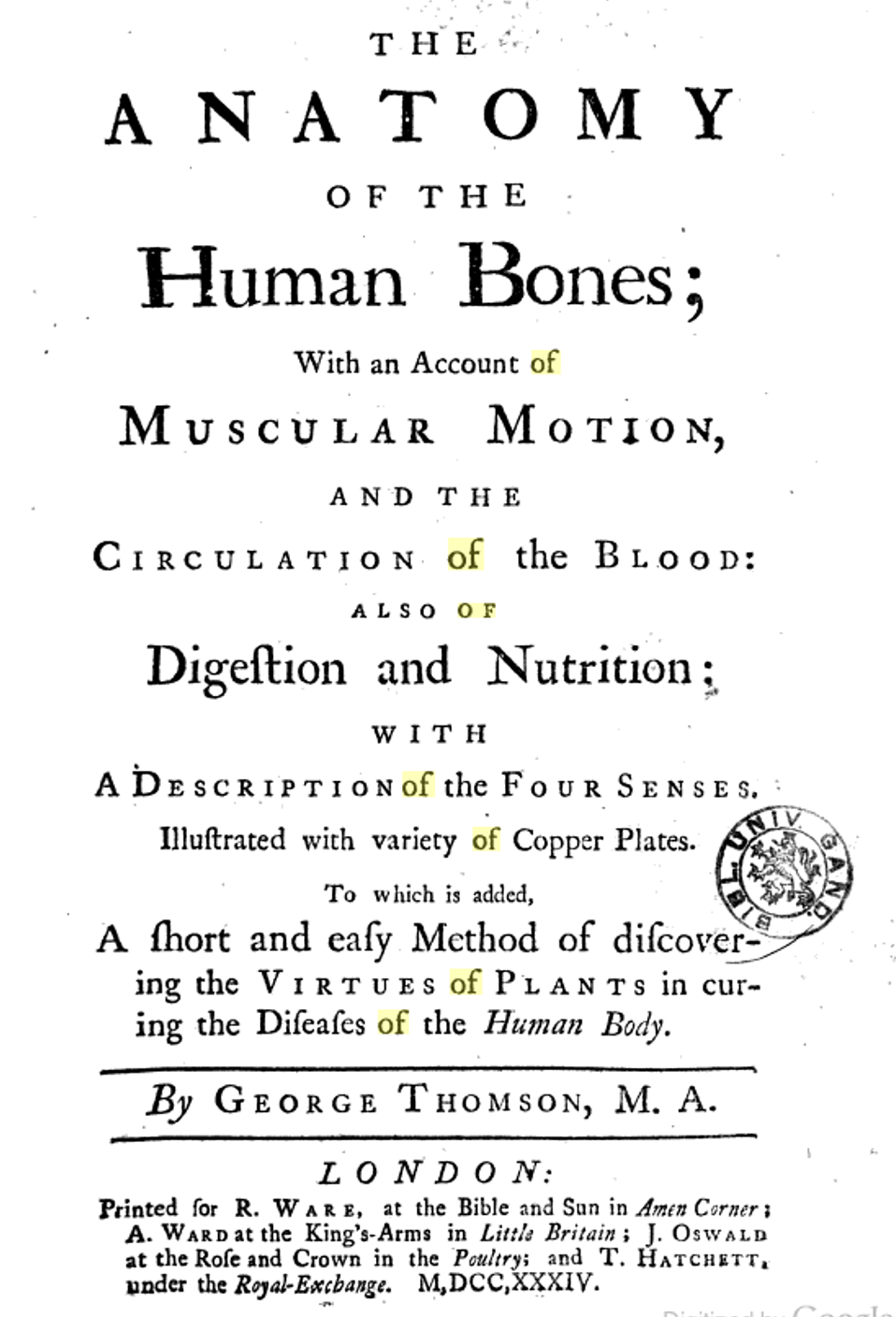
 The inner ear was believed to be air-filled. This was because cerebrospinal fluid pressure rapidly becomes negative after death. Disturbance of the stapes when you are trying to look inside the ear allows air to be rapidly drawn into the labyrinth as the perilymph is sucked into the cranium through the cochlear aqueduct. By the time you could look inside, it was air-filled.
Sound was thought to be transmitted to all the organs inside the ear. The entire labyrinth was regarded as “the immediate organ of hearing”.
 Thomson made the argument that there is no spiral cochlea in birds or fish, so the semicircular canals are part of the “immediate organ of hearing”. Furthermore the semicircular canals act in a way that the “impression of sounds increases and fortifies itself” as it passes through the ear.
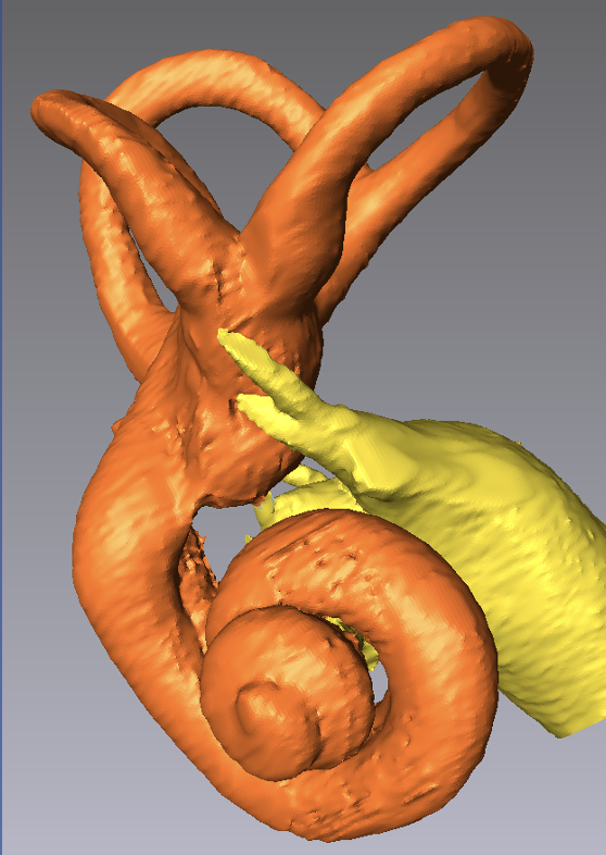
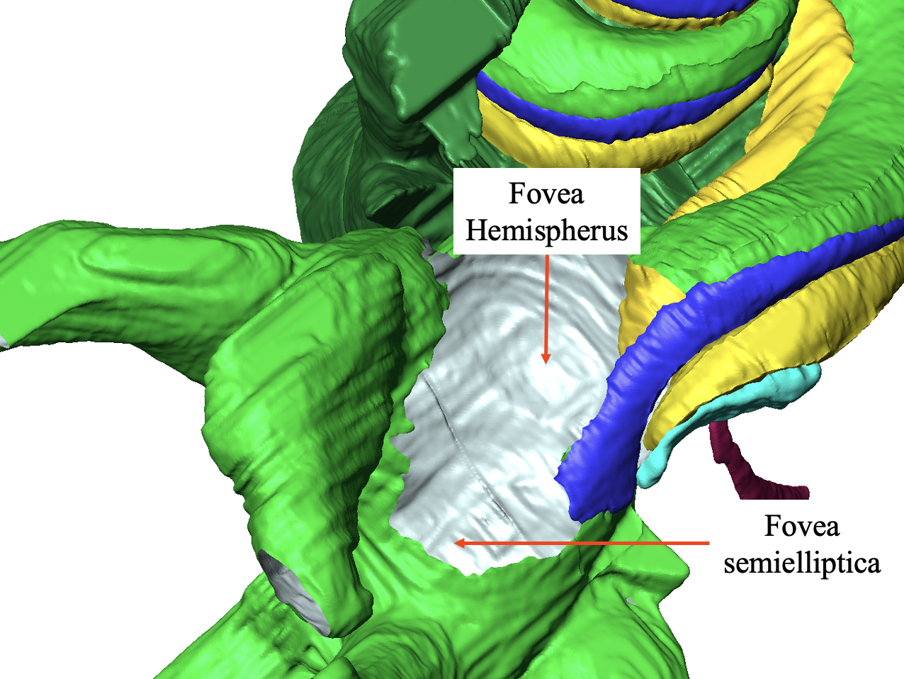
At this point, just consider what was seen when the stapes was removed and you looked through into the vestibule The below figure shows a 3D reconstruction of the human ear with the stapes, surrounding bone and vestibular endolymphatic structures removed. The inside of the structure is shown silver. Without the soft tissues of the saccular and utricular maculae you would see depressions in the bone underlying the structures when the stapes was removed. These were regarded as mechanical structures to amplify/focus the sound and were accordingly given the terms “fovea hemisherus” and “fovea semielliptica” (analogous to the fovea of the eye). With the stapes applying sounds directly above these structures it seemed logical that they were involved in the amplification of the sound.
Now let us consider the "distasteful" work of Flourens 1817, as described in an 1884 textbook by Burnett. Flourens was the first to suggest that disruption of the semicircular canals caused “peculiar disturbances of equilibrium of the body”
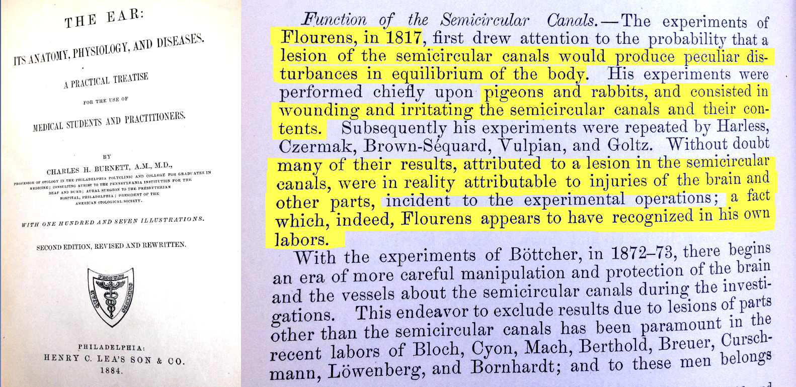 However, Flourens experiments were performed in conscious, un-anesthetized animals and involved drilling holes into the ear or brain and in some cases pulling on the nerves. Damage to one SCC was observed to cause the head to snap in a specific direction. Even then, they were regarded as unethical and were largely ignored by the medical and scientific communities.
In the Cyclopedia of Anatomy and Physiology 1839, edited by Todd, it is stated that the vestibule is part of the hearing mechanism 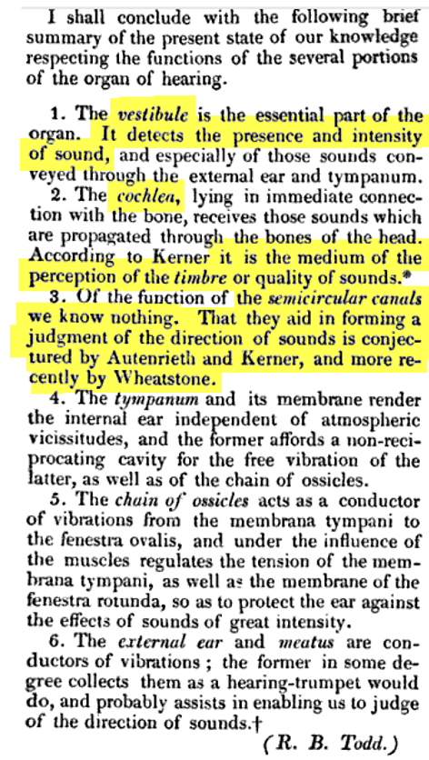
Todd further dismisses the work of Flourens 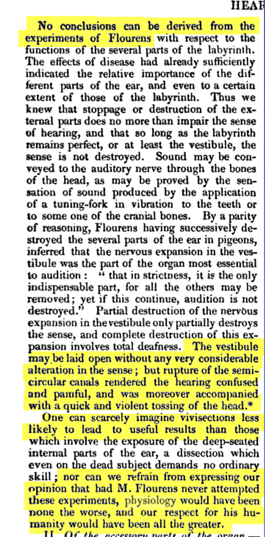
An anatomic textbook of the same year (1839) by Wistar and Horner shows a mid-modiolar section of the human cochlea in which a membrane divides the spiral tube into two compartments. It should be appreciated that histology of the ear was in its infancy and the fibrous basilar membrane was the main structure that was observed within. At this time the sensory organ and sensory cells were not visible (subsequently reported by Corti in 1851) as was the membrane forming a boundary between the endolymph space and scala vestibuli (reported by Reissner in 1854).
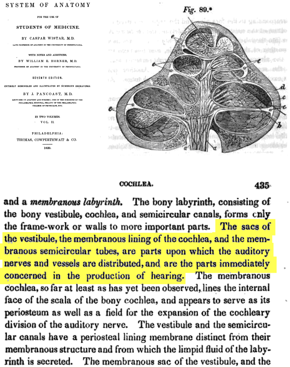
From the text It is apparent that both the vestibular stuctures and the cochlea are still believed to be involved only in hearing.
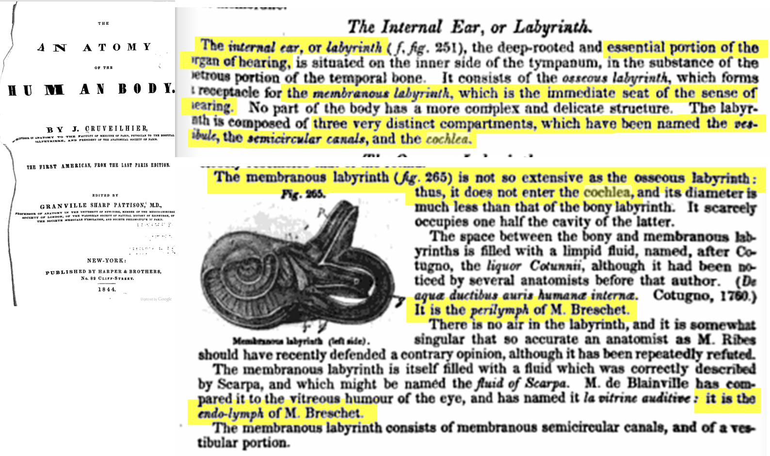
This was a period of rapid discovery and in 1844 a text by Cruveilhier identifies the membranous labyrinth containing endolymph (attributed to the French anatomist Breschet) within an outer compartment containing perilymph. The text emphasizes that there is no air in the labyrinth even though some anatomists (Ribes) still hold that view. The entire labyrinth remains the “organ of hearing”
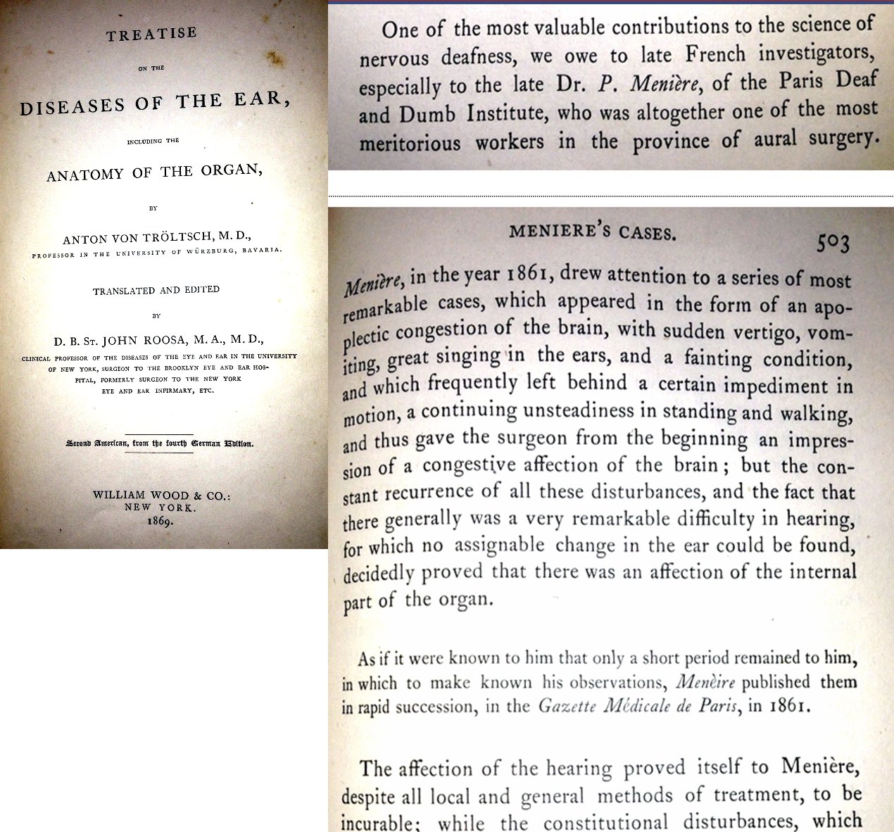
In 1861, just prior to his death in 1862, Prosper Menière published his paper attributing the dizziness of a patient to a pathology of the semicircular canals. From the above texts, this observation was presented to a medical and scientific community that believed the inner ear was only involved in hearing. Meniere's perspective was not well accepted.
From a translation by Politzer, 1902 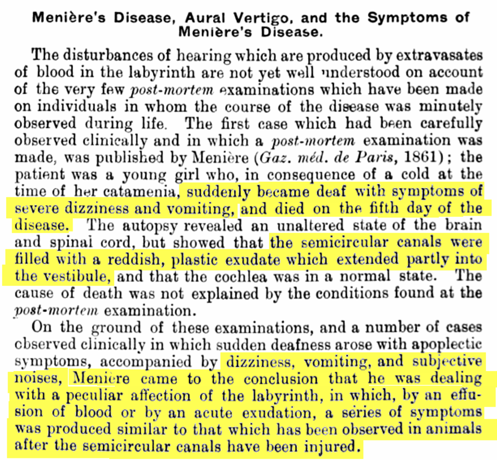
Although Meniere's conclusions were not initially accepted, some began to consider his observations. In 1869 Tröltsch presents Menière’s cases, with conjecture that “the seat of the disease” was the semicircular canals. Elsewhere in the text he also compares Menière’s observations with those of Flourens.
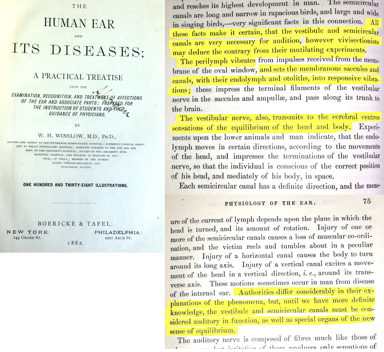
In 1882, Winslow shows some acceptance of the concept but suggests that vestibular structures “must be considered auditory in function, as well as special organs of the new sense of equilibrium”.
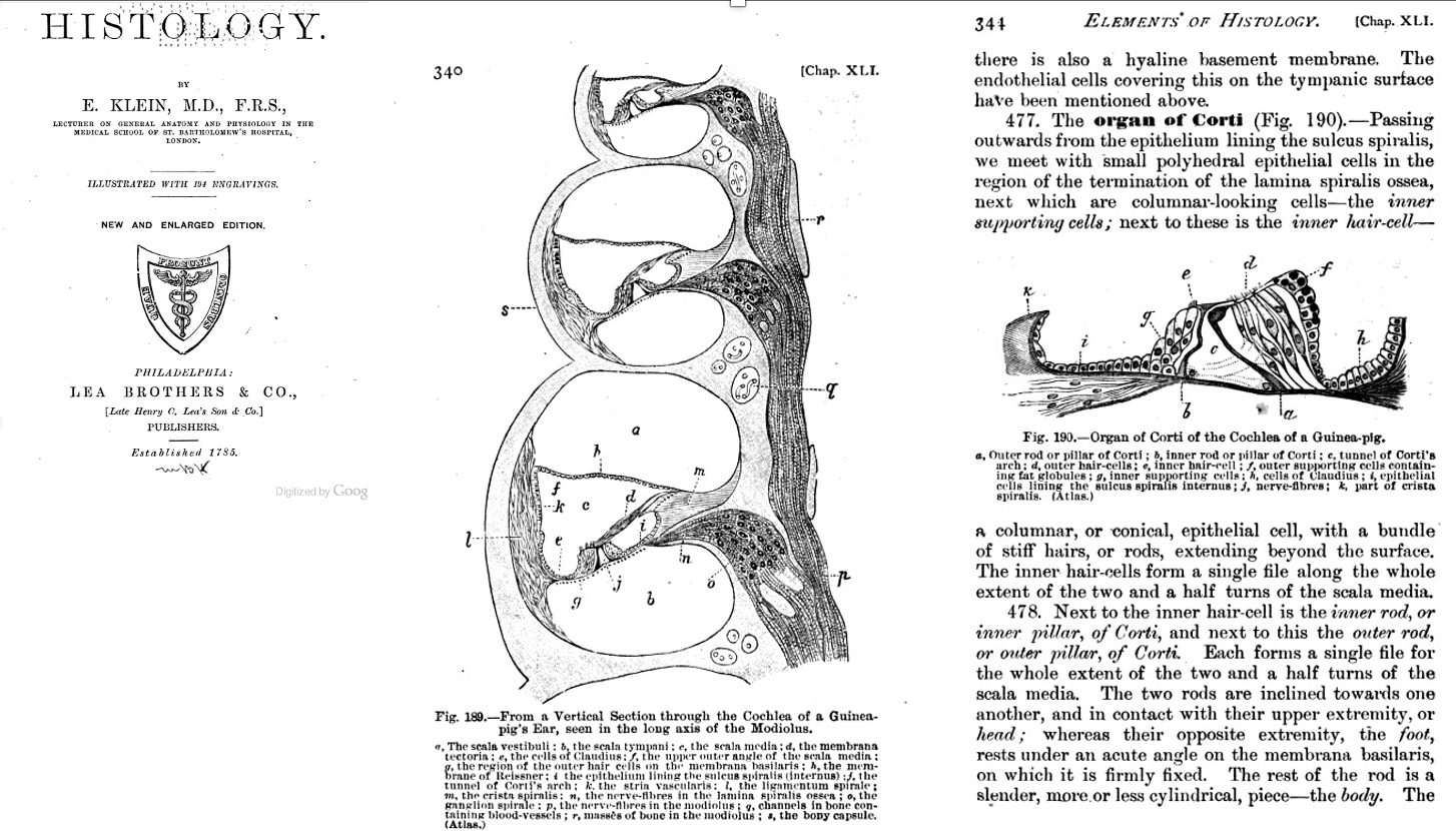
By 1899 more detailed anatomical descriptions of the cochlea had made it to the textbooks, with the organ of Corti, Reissner’s membrane, inner hair cells, outer hair cells and stria vascularis all identified.
One of the most reputable textbooks of the period, for which English translations are available, was assembled by Adam Politzer. In 1861 Politzer had spent a research fellowship in Paris, which involved visits to the Institute for Deaf-Mutes where Menière was the “physician in residence”. He was well aware of Menière’s concepts. There were two editions of Politzer’s texts, one in 1883 and a second in 1902. 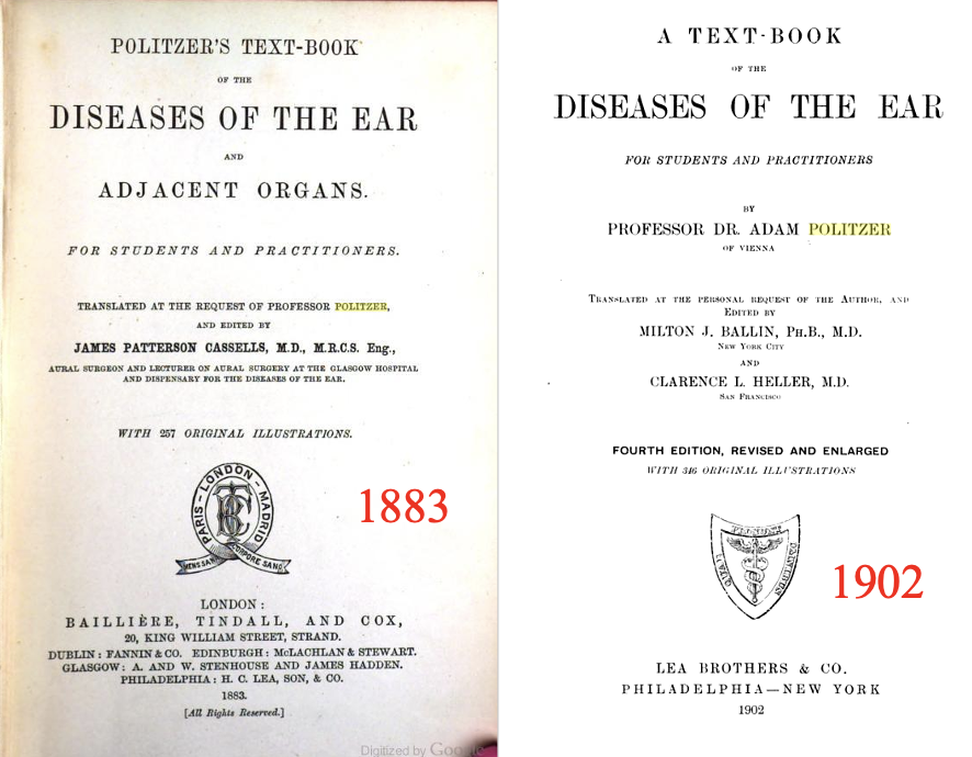
In the introductory paragraphs of both versions concerning the anatomical division of the ear, the labyrinth is included in the “sound-perceiving apparatus” with no mention of the involvement in equilibrium and balance. 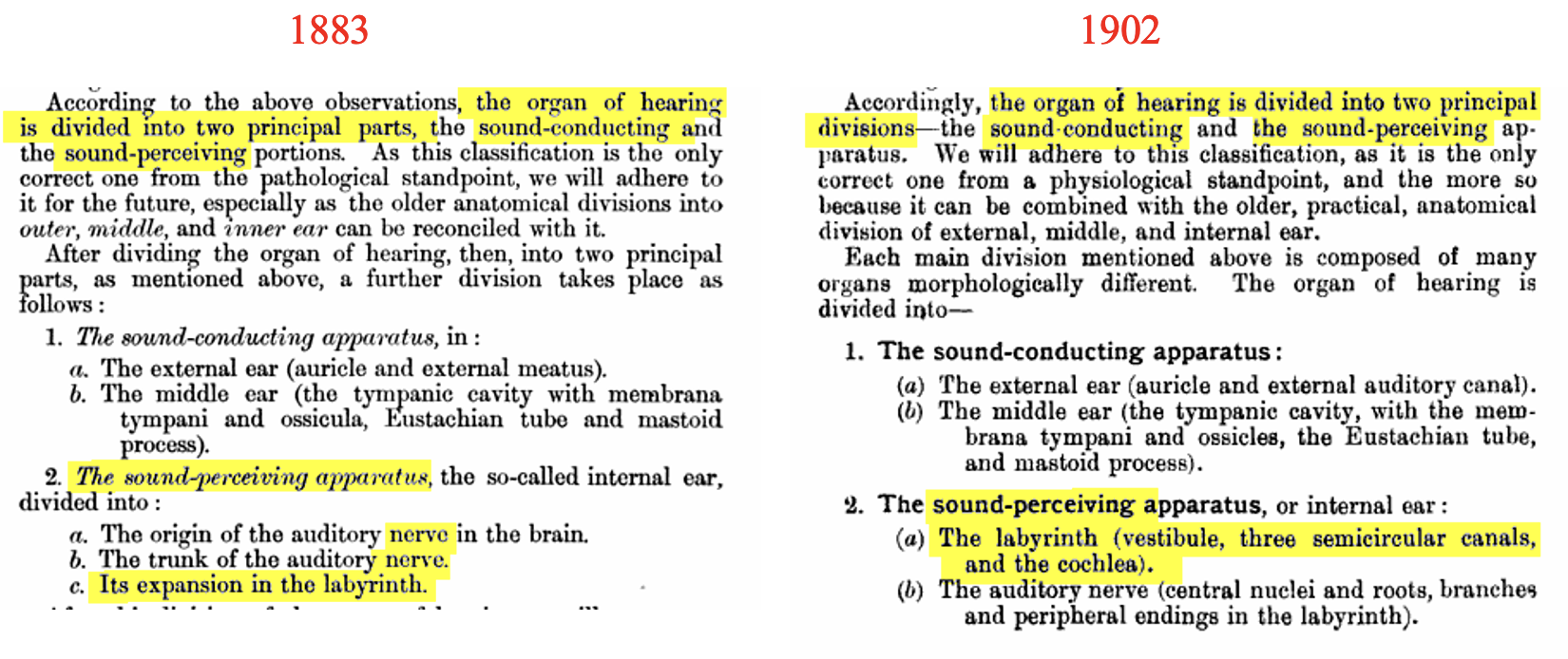
In the section relating to the physiology of the ear the 1883 version stated that the function of the vestibular ototliths was to damp the sound. By 1902, the vestibular apparatus was recognized, including the involvement in head motions and equilibrium. Furthermore the pioneering work of Flourens was now recognized.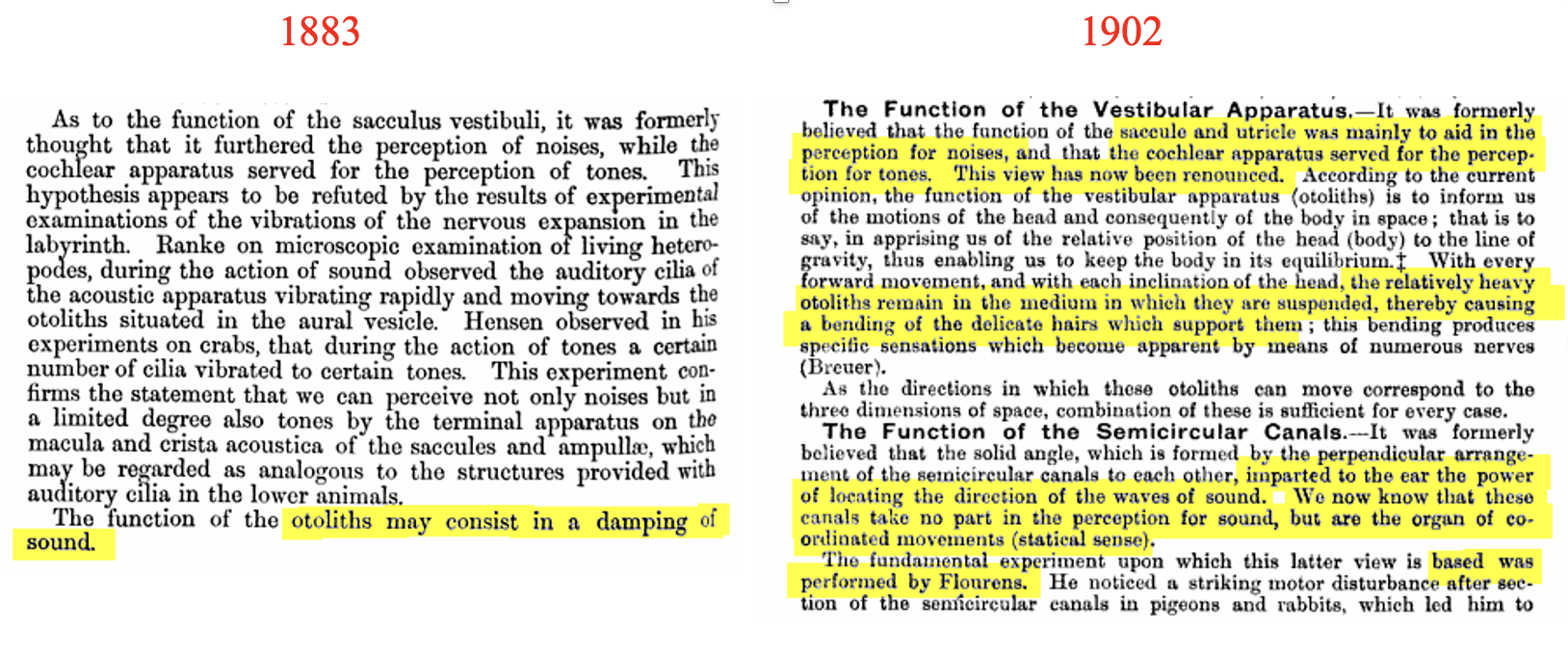 Concerning Meniere’s disease, the 1883 edition presented the disease as a brief subsection on the topic of haemorrhages into the labyrinth. In contrast the 1902 version included a detailed, comprehensive 14 page discussion of Menière’s disease. This publication played an important role in bringing Menière’s work to the attention of “mainstream” ENT physicians of the period
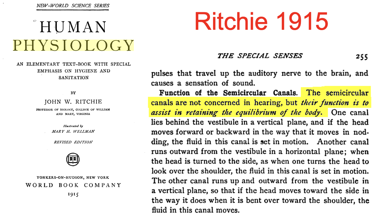
 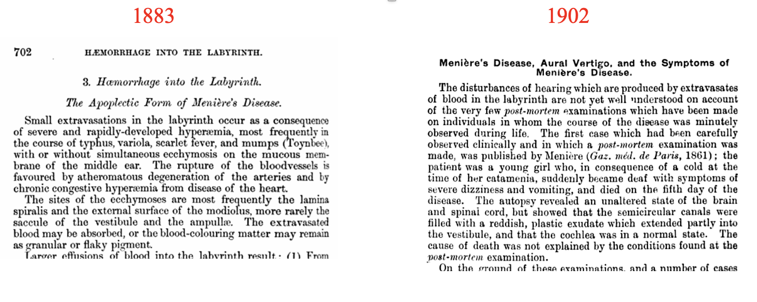 By 1915 most textbooks has incorporated the new thinking, that the vestibular system played a role in the equilibrium of the body.
But there were hold-outs. In “The Medical Record” in 1908, George Gould presented an 8 page diatribe against the entire concept of Menière’s disease and the “false and snapped-up theory that the labyrinth was the organ of equilibrium".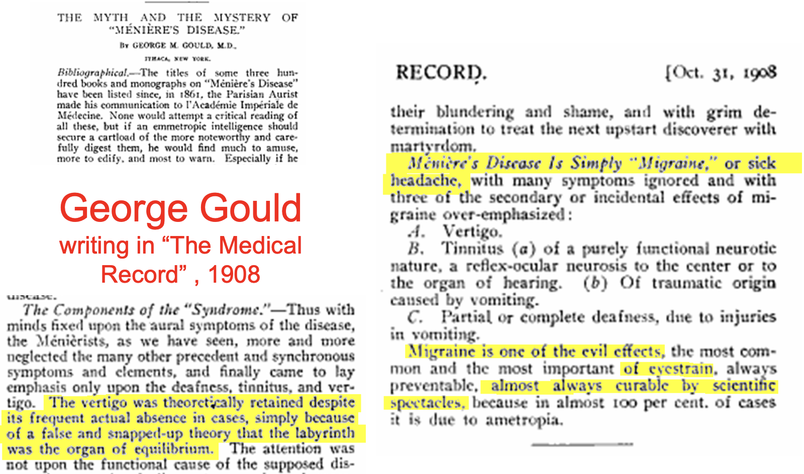
Conclusion:
Prosper Meniere’s contribution to the field was much greater than characterizing the medical condition now known as Meniere’s disease. The case presented in the 1861 article was important because it was a clear description of a balance disorder resulting from pathology of a semi-circular canal, presented by a reputable physician. It suggested the semicircular canals were primarily involved in balance and not in hearing, strongly in contrast to the prevailing scientific consensus. It took over 40 years for the truth to become widely accepted by the scientific and medical communities. Challenging the concensus is often a difficult battle.
But – the story doesn’t end there. Remember the 2010 Cummings textbook in which it was stated “The portion of the inner ear that deals with hearing is the cochlea and the portion of the inner ear that deals with balance is collectively known as the vestibular organs (semicircular canals, utricle and saccule).” This documents the current consensus.
However, the saccule has been shown to respond to acoustic stimulation and is the basis of the clinical VEMP response used to test saccular function. Similarly, SCC, saccule and utricle receptors have been shown to respond to acoustic clicks at above 60 dB SL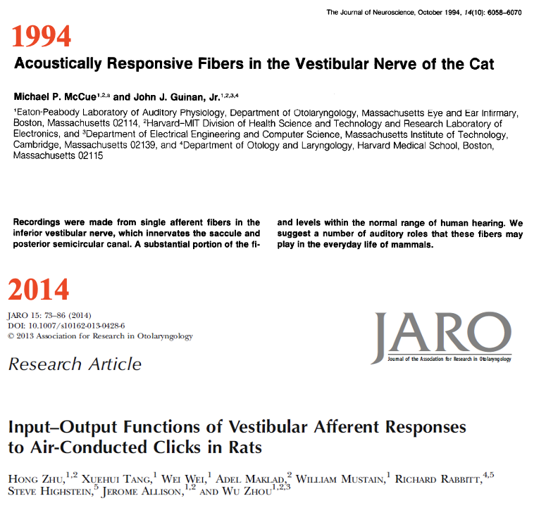
It is therefore likely that the entire ear responds to air-borne acoustic stimulation, especially for loud sounds. Meniere was correct in associating the vestibular organs with balance but this does not exclude the possibility that vestibular organs could serve a dual purpose.
Texts in which it is assumed that all vestibular function is related to balance and all cochlear function is related to hearing may be oversimplifying the physiology of the ear. Current consensus may be oversimplified.
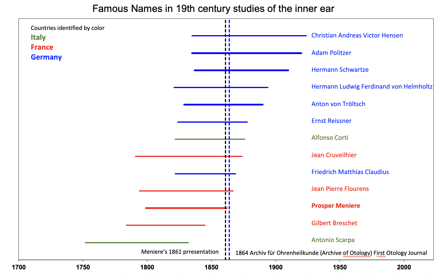
We finish with a timeline of some of the major contributors to our understanding of the inner ear. This period provided the foundation for what we know as the field of Otology today. The first otology journal, Archiv für Ohrenheilkunde (Archive of Otology) was published in Halle, Germany in 1864. The editors were Anton von Tröltsch, Hermann Schwartze (in Halle) and Adam Politzer in Wien. The journal subsequently became the European Archives of Oto-rhino-laryngology.